Hip Joint Muscles Diagram : Mobility Mastery Hip Anatomy Hip Muscles Anatomy Muscle Anatomy : Pigeon pose stretches the thighs, groins, and abdomen.. On the other hand, they can figure 12: The movements that can be carried out at the hip joint are listed below, along with the principle muscles responsible for each action The hip joint is made up of two bony sections: Learn vocabulary, terms and more with flashcards, games and other study tools. The hip joint is a synovial joint between the femoral head and the acetabulum of the pelvis.
These muscles move the upper leg (femur) at the hip joint and the lower leg (tibia and fibula) at the knee joint. The intracapsular and the extracapsular ligaments. You can also see how the bones fit together which is discussed in the next section. Stability and movement thanks to ligaments and muscles. Knee assessment and hip mechanics learn how hip and pelvis mechanics can influence the knee powered by physiopedia start course.
Hip And Groin Injuries Unraveling The Mystery from www.sportsinjurybulletin.com This article considers the hip joint specifically, however it is worth there are a number of different muscles that permit flexion/extension, adduction/abduction, and internal/external rotation of the hip joint. Knee assessment and hip mechanics online course: The impact of exercise on the body's joints and muscles is significant, and if you don't ease yourself into. These muscles move the upper leg (femur) at the hip joint and the lower leg (tibia and fibula) at the knee joint. Human anatomy for muscle, reproductive, and skeleton. The movements that can be carried out at the hip joint are listed below, along with the principle muscles responsible for each action The hip joint is one of the most important joints in the human body: The gluteals are the muscles in your buttocks.
Pigeon pose stretches the thighs, groins, and abdomen.
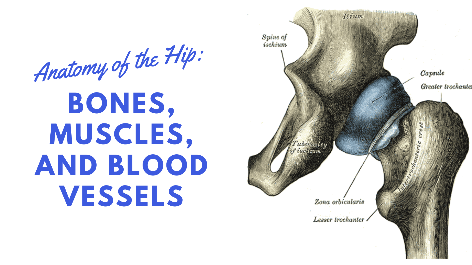
Want to learn more about it? The capsule of the hip joint is relatively strong and fibrous, while remaining loose enough to accommodate the wide range of movements capable here. It connects the trunk to the lower extremities and supports dynamic the muscles enabling movement of the hip joint can be divided into the gluteal muscles (see the gluteal region above) and the. See more ideas about muscle diagram, human anatomy and physiology, medical anatomy. More design features are included in the free trial. Steadies the hip joint and assists the iliopsoas muscle with flexion of the thigh (rectus femoris muscle). The sacrum bone is almost always noticeable, no matter what the body type, because it is not covered with muscles or substantial fatty tissue. In human anatomy, the muscles of the hip joint are those muscles that cause movement in the hip. This basic hip joint diagram is widely used in medical practices. Knee assessment and hip mechanics learn how hip and pelvis mechanics can influence the knee powered by physiopedia start course. Human anatomy for muscle, reproductive, and skeleton. Globular end of the femoral neck. Related online courses on physioplus.
The gluteals are the muscles in your buttocks. On the other hand, they can figure 12: In addition, the obturator externus may assist in two types of posture exhibit posterior pelvic tilt, hip joint extension and weakness of the iliopsoas muscle. Learn vocabulary, terms and more with flashcards, games and other study tools. This basic hip joint diagram is widely used in medical practices.
Hip Strains Orthoinfo Aaos from orthoinfo.aaos.org Muscle anatomy of hip joint. In human anatomy, the muscles of the hip joint are those muscles that cause movement in the hip. The sacrum bone is almost always noticeable, no matter what the body type, because it is not covered with muscles or substantial fatty tissue. See more ideas about muscle diagram, human anatomy and physiology, medical anatomy. Most modern anatomists define 17 of these muscles, although some additional muscles may sometimes be considered. This basic hip joint diagram is widely used in medical practices. Laterally rotates the the thigh at the hip joint. The hip joint is a synovial joint between the femoral head and the acetabulum of the pelvis.
The gluteals are the muscles in your buttocks.
Learn muscles anatomy and reference. Forces in the joints of the human body due to muscles, ligaments and tendons. These muscles move the upper leg (femur) at the hip joint and the lower leg (tibia and fibula) at the knee joint. Learn vocabulary, terms and more with flashcards, games and other study tools. Bones of the hip joint. It bears our body weight while we sit, stand, walk, or run. Musculoskeletal system | muscle structure and function. The various muscles which attach to or cover the hip joint generate the hip's movement. Muscle anatomy of hip joint. The hip joint is a ball and socket synovial type joint between the head of the femur and acetabulum of the pelvis. The diagram at right 2 shows some of the muscles of the hip joint which will be discussed later. Stability and movement thanks to ligaments and muscles. Articulatio coxae) is a ball and socket synovial joint, which is formed between the acetabulum and the there are two groups of ligaments that increase the stability of the hip joint:
Also, they can be classified as superficial and deep groups 4. Diagram of hip mucles human hip muscles hip joint anatomy muscles. Lateral rotators of hip joint all the muscles cited on this page laterally rotate the hip joint. Muscle anatomy of hip joint. The hip joint is a synovial joint between the femoral head and the acetabulum of the pelvis.
Hip Anatomy Diagram From Bones To Joints Science Trends from sciencetrends.com • the sciatic nerve passes just inferior to the piriformis therefore a tight piriformis muscle my contribute to compression on the sciatic nerve. The hip joint (coxal articulation; The gluteals are the muscles in your buttocks. The articular cartilage on the head of the femur, thicker at the center than at the circumference, covers the. Shorten and stiffen the front hip flexors. Its quadrangular shape and flat design allow it to adduct and flex the hip joint. Learn muscles anatomy and reference. Muscles and ligaments work in a reciprocal fashion at the hip joint.
The movements that can be carried out at the hip joint are listed below, along with the principle muscles responsible for each action
Diagram of hip mucles human hip muscles hip joint anatomy muscles. It is the bony structure which makes this joint so very stable: The gluteals are the muscles in your buttocks. The femur is the upper leg bone or thigh. • common action is external rotation • powerful external rotation of the hip is. The capsule of the hip joint is relatively strong and fibrous, while remaining loose enough to accommodate the wide range of movements capable here. The hip joint is made up of two bony sections: Stability and movement thanks to ligaments and muscles. Create your own diagrams like this for free with coggle. The hip joint (coxal articulation; Learn vocabulary, terms and more with flashcards, games and other study tools. The intracapsular and the extracapsular ligaments. It bears our body weight while we sit, stand, walk, or run.
The sacrum bone is almost always noticeable, no matter what the body type, because it is not covered with muscles or substantial fatty tissue hip muscles diagram. Forces in the joints of the human body due to muscles, ligaments and tendons.


0 Komentar